
Modern technologies in neurosurgery
An integrated technology pathway for diagnostics, surgical planning, navigation, and intraoperative control.
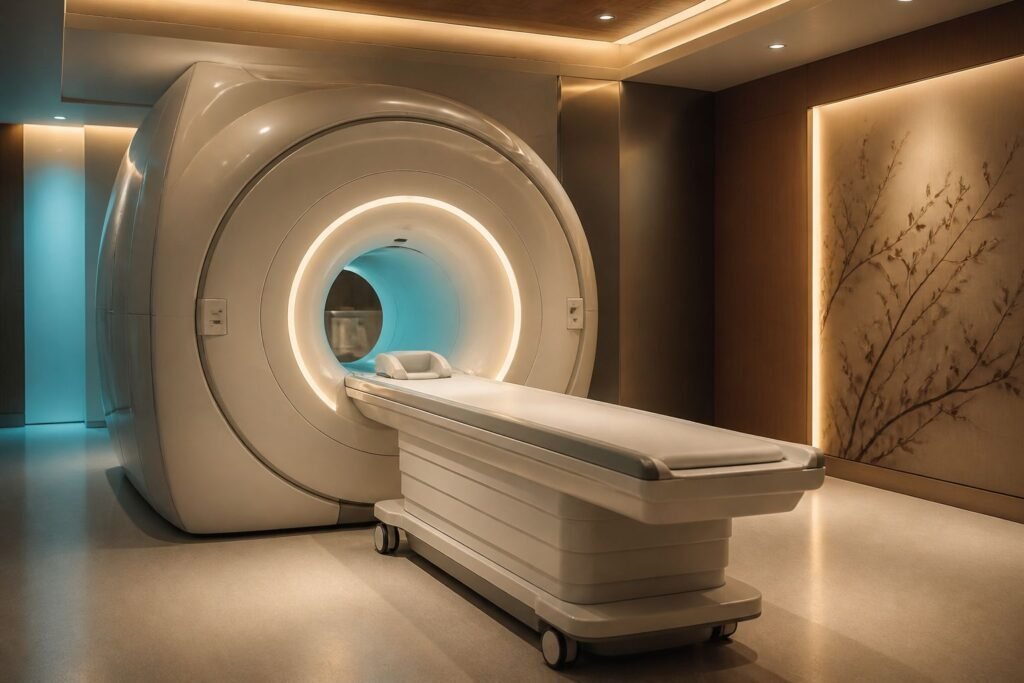
3-tesla magnetic resonance imaging
GE SIGNA Hero 3T
High-detail MRI for assessing the brain, spine, and vascular structures. The images support a more precise diagnosis and individualized surgical planning.
Key advantages and use
- 3T magnetic field and high spatial detail
- Multiparametric studies of the brain and spine
- 70 cm wide bore for patient comfort
- Anatomical information needed for surgical planning
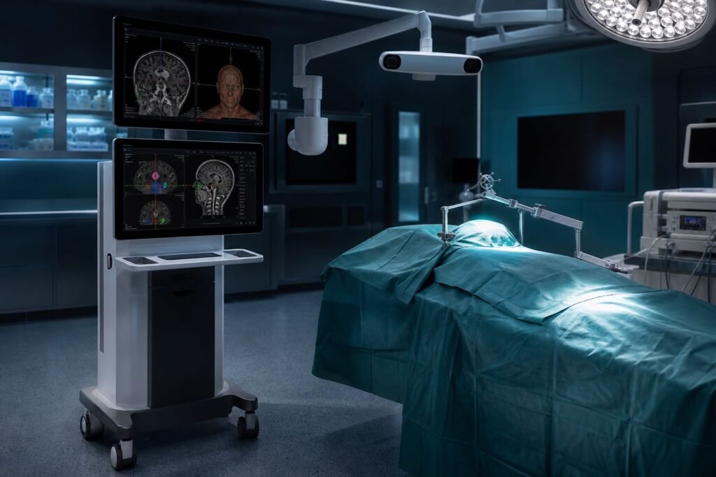
Surgical neuronavigation system
Navient
Links preoperative CT/MRI data to the patient’s anatomy and shows the surgeon the instrument position in real time. Used for precise trajectory planning and orientation to the anatomical target.
Key advantages and use
- Real-time display of instrument spatial position
- Support for cranial, spinal, and biopsy procedures
- Use of DICOM images and image fusion
- Preoperative planning of trajectory and target
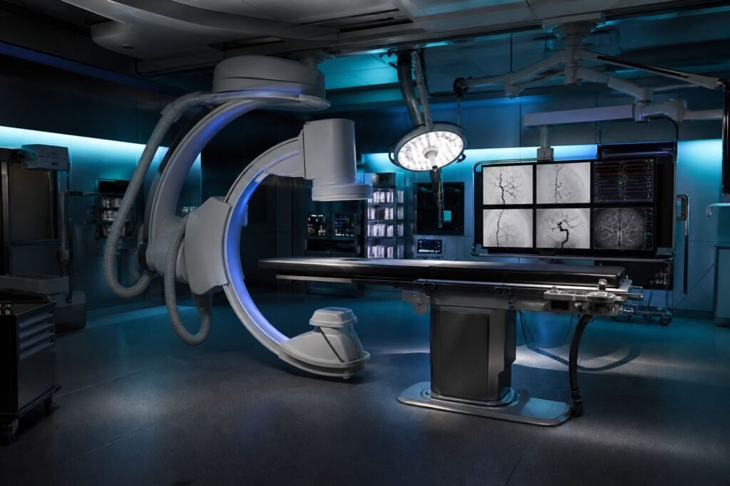
Angiographic and hybrid operating system
GE HealthCare Allia IGS 5
An interventional imaging system for visualizing vascular anatomy and guiding endovascular procedures. In neurosurgery it is used, among other things, for cerebral angiography and corresponding minimally invasive procedures.
Key advantages and use
- Real-time fluoroscopic and angiographic visualization
- Control of endovascular instrument movement
- 3D vascular reconstruction in the corresponding configuration
- Workflow integration in a hybrid operating environment
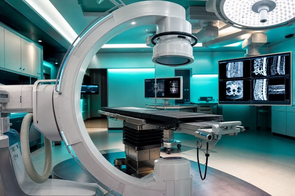
Intraoperative 3D C-arm
GE OEC 3D
Creates 2D fluoroscopic and three-dimensional images during surgery. In spine surgery it can be used to check anatomy and the position of implants and fixation hardware.
Key advantages and use
- Intraoperative 3D imaging
- 3D acquisition volume of 19 × 19 × 19 cm
- 2D fluoroscopy and CT-like multiplanar views
- Intraoperative control of implants and anatomical alignment
From diagnosis to intraoperative control
SIGNA Hero 3T provides detailed diagnostic information for surgical planning; Navient turns preoperative images into real-time surgical orientation; Allia IGS 5 delivers angiographic and endovascular visualization; OEC 3D adds three-dimensional control during surgery. Together the four systems connect four stages of patient care and do not replace the surgeon’s clinical judgment.
Availability of specific features depends on the installed configuration, software options, and clinical protocol.